
Back Muscle Diagrams 101 Diagrams
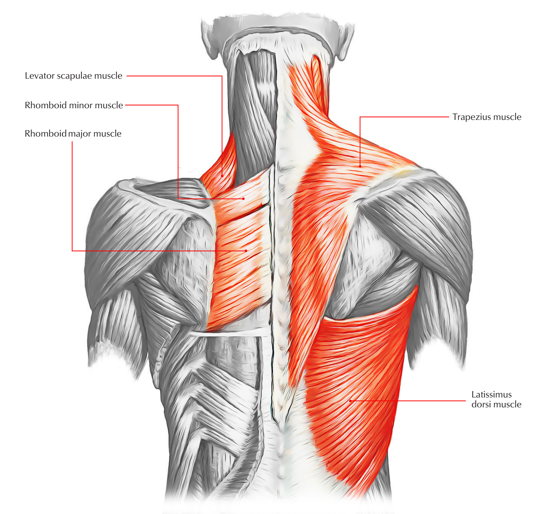
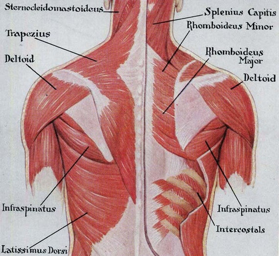
The first subgroup consists of two superficial muscles: the trapezius and latissimus dorsi. The trapezius is a large triangular muscle that covers the posterior aspect of the neck and the superior half of the back. There are two trapezius muscles in the back, which when seen together, look like a trapezium. Proximally, the trapezius originates.
88+ Muscles Of The Back l2sanpiero
The human back, also called the dorsum (pl.: dorsa), is the large posterior area of the human body, rising from the top of the buttocks to the back of the neck. It is the surface of the body opposite from the chest and the abdomen.The vertebral column runs the length of the back and creates a central area of recession. The breadth of the back is created by the shoulders at the top and the.

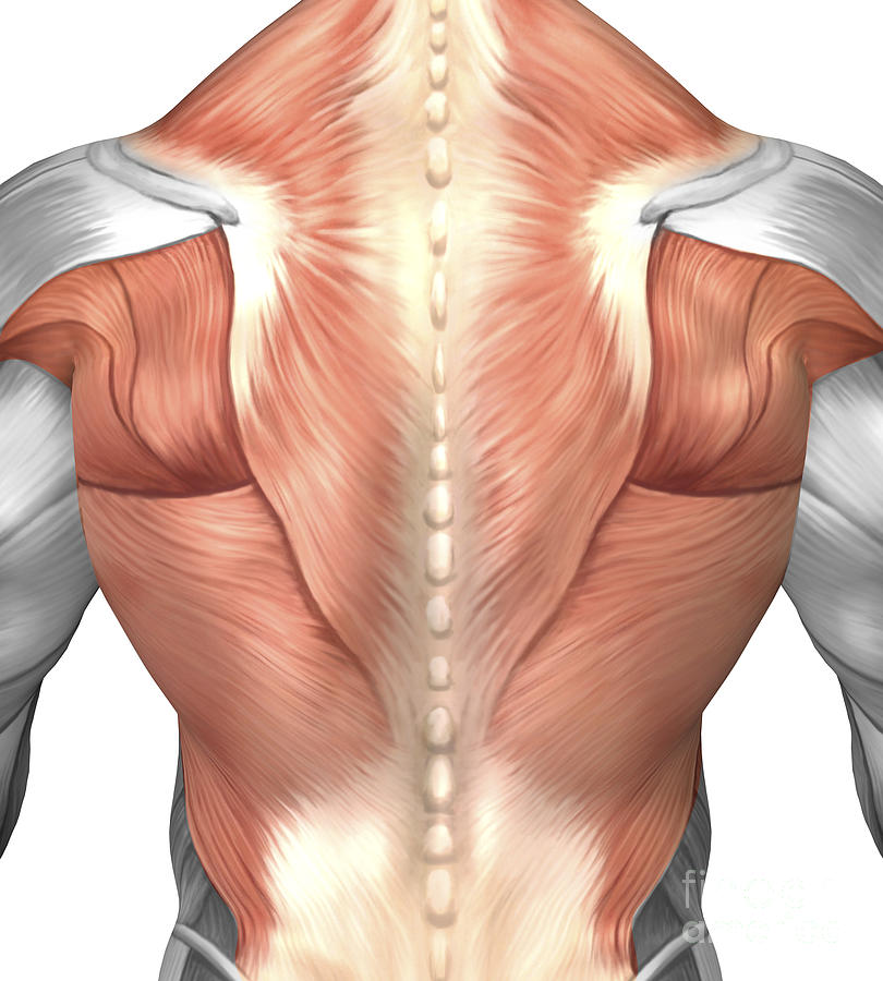
Beautiful illustration of the deep and superficial musculature of the back. This anatomical
The back is a key topographical region of the body, with crucial importance for posture, locomotion, and upper and lower limb movements. [1] The spine, located in the midline, divides the body into unequal anterior and posterior segments. In the posterior segment, the body area between the neck and gluteal regions is defined as the back region.
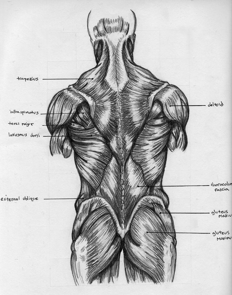
Back Muscles Chart by BadFish81 on DeviantArt
Human Anatomy - Back View of Muscles. Click on the labels below to find out more about your muscles. More human anatomy diagrams: front view of muscles, skeleton, organs, nervous system. Flex some.
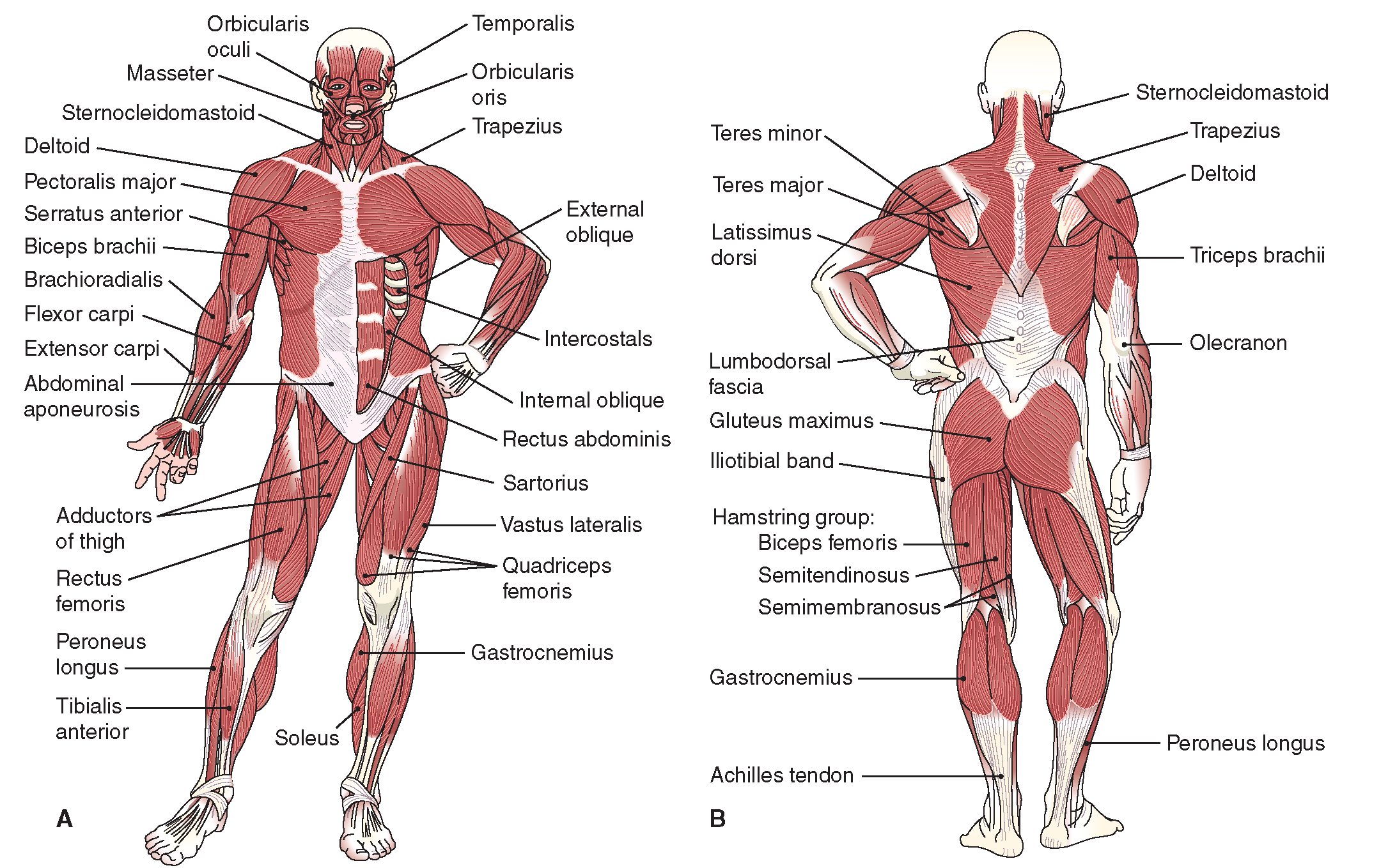
The Musculoskeletal System (Structure and Function) (Nursing) Part 4
Browse Getty Images' premium collection of high-quality, authentic Human Anatomy Organs Back View stock photos, royalty-free images, and pictures. Human Anatomy Organs Back View stock photos are available in a variety of sizes and formats to fit your needs.. engraving human body with muscles 1851 - human anatomy organs back view stock.
Back Muscles Diagram Unlabeled / Back Muscles 28 Major Muscles Of The Back Earth S Lab Human
The muscles of the back categorize into three groups. The intrinsic or deep muscles are those muscles that fuse with the vertebral column. The second group is the superficial muscles, which help with shoulder and neck movements. The final group is the intermediate muscles, which help with the movement of the thoracic cage. Only the intrinsic muscles are considered true back muscles.

bodyman Full back muscles
Summary. The back consists of the spine, spinal cord, muscles, ligaments, and nerves. These structures work together to support the body, enable a range of movements, and send messages from the.
Male Muscle Anatomy Of The Human Back Digital Art by Stocktrek Images Pixels
The back is the body region between the neck and the gluteal regions. It comprises the vertebral column (spine) and two compartments of back muscles; extrinsic and intrinsic. The back functions are many, such as to house and protect the spinal cord, hold the body and head upright, and adjust the movements of the upper and lower limbs.

Anatomy of back muscles Diagram Quizlet
The back comprises the dorsal part of the neck and the torso (dorsal body cavity) from the occipital bone to the top of the tailbone.The muscles of the back can be divided in three main groups according to their anatomical position and function. The superficial muscles participate in the movements of the upper limb, the intermediate muscles support the respiratory function, and the deep.
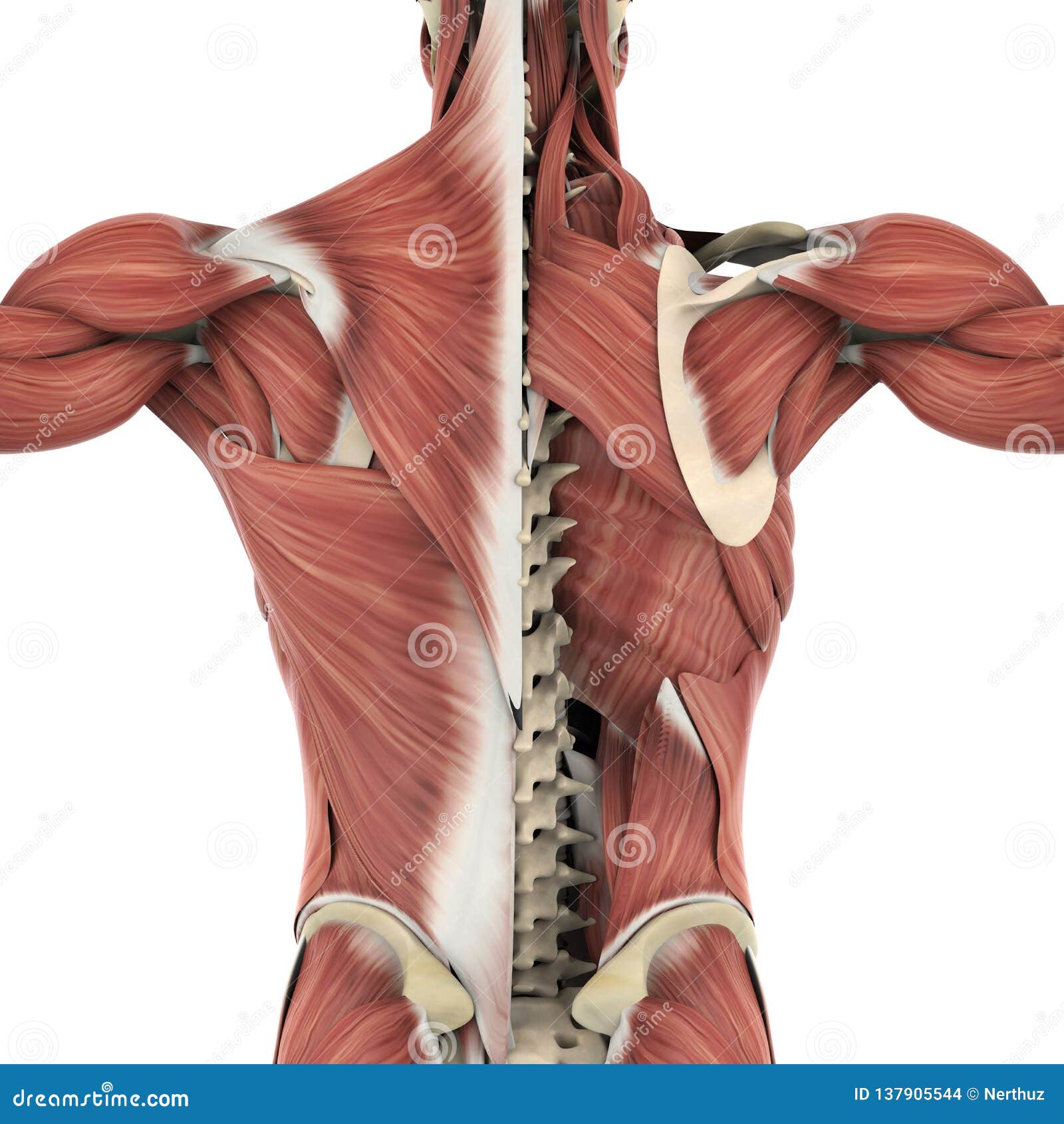
Muscles of the Back Anatomy Stock Illustration Illustration of isolated, muscle 137905544
ISSN 2534-5079. This human anatomy module is composed of diagrams, illustrations and 3D views of the back, cervical, thoracic and lumbar spinal areas as well as the various vertebrae. It contains the osteology, arthrology and myology of the spine and back. It is particularly interesting for physiotherapists, osteopaths, rheumatologists.

Muscle Anatomy Posterior Human Anatomy Muscles Of The Back Muscular System Posterior View
Flashcard: Muscles back view. This is only a glimpse of what you can do in Complete Anatomy. This state of the art digital atlas of 3D imagery allows you to explore the human body on your tablet and phone - layer by layer, and from every possible angle. So aside from testing your anatomical knowledge, the app helps you to fully understand the.

Superficial Back Muscles, 3 layers Diagram Quizlet
Your back consists of a complex array of bones, discs, nerves, joints, and muscles. The muscles of your back support your spine, attach your pelvis and shoulders to your trunk, and provide mobility and stability to your trunk and spine. The anatomy of your back muscles can be complex. There are several different layers of muscles in your back.
Muscles Back Posterior Human Anatomy Vintage Medical Chart
The muscles of the back are a group of strong, paired muscles that lie on the posterior aspect of the trunk. They provide movements of the spine, stability to the trunk, as well as the coordination between the movements of the limbs and trunk. The extrinsic (superficial) back muscles, which lie most superficially on the back.

Muscle Facts Human Back Muscles DK Find Out
The muscles of the back can be arranged into 3 categories based on their location: superficial back muscles, intermediate back muscles and intrinsic back muscles.The intrinsic muscles are named as such because their embryological development begins in the back, oppose to the superficial and intermediate back muscles which develop elsewhere and are therefore classed as extrinsic muscles.

Human Anatomy Back . Human Anatomy Back Anatomy Human Muscles Back Template Medical
This video provides an overview of the muscles of the back (superficial, intermediate and deep) using high-quality 3D anatomy models and expert narration fro.

Muscles Diagrams Diagram of muscles and anatomy charts HubPages
Latissimus dorsi (lats), the largest muscle in the upper part of your body. It starts below your shoulder blades and extends to your spine in the lower part of your back. Levator scapulae, a smaller muscle that starts at the side of your neck and extends to the scapula (shoulder blade). Rhomboids, two muscles that connect the scapula to the spine.